-
G6D-311
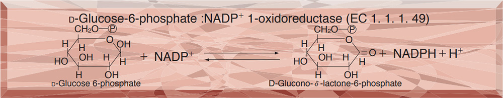
GLUCOSE-6-PHOSPHATE DEHYDROGENASE from Leuconostoc mesenteroides
PREPARATION and SPECIFICATION
| Appearance | White amorphous powder, lyophilized | |
|---|---|---|
| Activity | GradeⅢ 400U/mg-solid or more (NAD+) | |
| Contaminants | Creatine phosphokinase | ≤1×10⁻³% |
| Phosphoglucomutase | ≤1×10⁻³% | |
| 6-Phosphogluconate dehydrogenase | ≤5×10⁻³% | |
| Phosphoglucose isomerase | ≤1×10⁻²% | |
| Glutathione reductase | ≤1×10⁻³% | |
| Hexokinase | ≤1×10⁻²% | |
| Myokinase | ≤1×10⁻²% | |
| NADH oxidase | ≤1×10⁻²% | |
| NADPH oxidase | ≤1×10⁻²% | |
PROPERTIES
| Stability | Stable at −20℃ for at least one year(Fig.1) | |
|---|---|---|
| Stable at 5℃ for at least 6 months (liquid form)(Fig.3) | ||
| Molecular weight | 104,000(two subunits of approx. 55,000) 1,2) | |
| Isoelectric point | 4.6 2) | |
| Michaelis constants 2) | NAD+ linked | 1.06×10⁻⁴M (NAD+), 5.27×10⁻⁵M (G-6-P) |
| NADP+ linked | 5.69×10⁻⁶M (NADP+), 8.1×10⁻⁵M (G-6-P) | |
| Structure | Neither cysteine nor cystine residues is present in the enzyme molecule 1) and essential lysine is indicated to be at active site. 3) | |
| Inhibitors | Acyl-CoA,4) ATP,4) mental ions etc. (Table 1) | |
| Optimum pH | 7.8(Fig.4) | |
| Optimum temperature | 50℃(Fig.5) | |
| pH Stability | pH 5.5−7.5 (30℃, 17hr)(Fig.6) | |
| Thermal stability | below 37℃ (pH 8.0, 30min)(Fig.7) | |
| Substrate specificity | Either NAD+ or NADP+ serves as coenzyme, the reaction velocity with NAD+ being approximately 1.8 times greater than with NADP.+5) DGlucose-6-phosphate is a preferential substrate for the enzyme, although D-glucose reacts slowly.6) Fructose-6-phosphate, fructose1, 6-diphoshate and ribose-5-phosphate are not considered to be substrates.7) | |
APPLICATIONS
This enzyme is useful for enzymatic determation of NAD+(NADP+) and G-6-P, and activities of phosphoglucose isomerase, phosphoglucomutase and hexokinase. This enzyme is also used for enzymatic determination of glucose when coupled with hexokinase (HXK-311).
ASSAY
Principle

The appearance of NADH is measured at 340nm by spectrophotometry.
Unit definition
One unit causes the formation of one micromole of NADH per minute under the conditions described below.
Method
Reagents
| A. Tris-HCl buffer, pH 7.8 | 55mM (containing 3.3mM magnesium chloride) | |
|---|---|---|
| B. NAD+ solution | 60mM (Should be prepared fresh) | |
| C. G-6-P solution | 0.1M glucose-6-phosphate (should be prepared fresh) | |
| D. Enzyme diluent | 5mM Tris-HCl buffer, pH 7.5, containing 0.1% of bovine serum albumin. | |
Procedure
1. Prepare the following reaction mixture in a cuvette (d=1.0cm) and equilibrate at 30℃ for about 5 minutes.
| 2.7ml | Tris-HCl buffer, pH 7.8 | (A) |
| 0.1ml | NAD+ solution | (B) |
| 0.1ml | G・6・P solution | (C) |
| Concentration in assay mixture | |
|---|---|
| Tris-HCl buffer | 50 mM |
| G-6-P | 3.3 mM |
| NAD+ | 2.0 mM |
| MgCl2 | 3.0 mM |
| BSA | 33μg/ml |
2. Add 0.1ml of the enzyme solution* and mix by gentle inversion.
3. Record the increase in optical density at 340nm against water for 4 to 5 minutes in a spectrophotometer thermostated at 30℃ and calculate the ΔOD per minute from the initial linear portion of the curve (ΔOD test).
At the same time, measure the blank rate (ΔOD blank) by using the same method as the test except that the enzyme diluent is added instead of the enzyme solution.
*Dissolve the enzyme preparation in ice-cold enzyme diluent (D) and dilute to 0.05−0.20U/ml with the same buffer, immediately before the assay.
Calculation
Activity can be calculated by using the following formula :
Volume activity (U/ml) =
-
ΔOD/min (ΔOD test−ΔOD blank)×Vt×df
6.22×1.0×Vs
= ΔOD/min×4.82×df
Weight activity (U/mg) = (U/ml)×1/C
| Vt | : Total volume (3.0ml) |
| Vs | : Sample volume (0.1ml) |
| 6.22 | : Millimolar extinction coefficient of NADH (cm2/micromole) |
| 1.0 | : Light path length (cm) |
| df | : Dilution facter |
| C | : Enzyme concentration in dissolution (c mg/ml) |
REFERENCES
1) A.Ishaque,M.Mihausen and H.R.Levy; Biochem. Biophys. Res. Comm., 59, 894 (1974).
2) C. Olive, M.E. Geroch and H.R.Levy; J.Biol.Chem., 246, 2043 (1971).
3) M.Milhausen and H.R. Levy; Eur.J.Biochem., 50, 453 (1975).
4) E.L.Coe and L.-H.Hsu; Biochem. Biophys. Res. Comm., 53, 66 (1973).
5) C.Olive and H.R. Levy; Biochem., 6, 730 (1967).
6) R.P.Metzger, S.A. Metzger and R.L. Parsons; Arch Biochem. Biophys., 149, 102 (1972).
7) Methods in Enzymology, Vol, 1, p328 (S.P.Colowick and N.O.Kapalan,eds.), Academic Press, New York (1955).
Table 1. Effect of Various Chemicals on Glucose-6-phosphate dehydrogenase
[The enzyme dissolved in 50mM Tris-HCl buffer,pH 7.5 (5.25U/ml) was incubated with each chemcal for 1hr at 30℃.]
-
Chemical Concn.(mM) Residual
activity(%)None - 100 Metal salt 2.0 AgNO3 86 Ba(OAc)2 51 CaCl2 90 Cd(OAc)2 74 CoCl2 80 CuSO4 66 FeCl3 0 FeSO4 1 HgCl2 84 MgCl2 90 MnCl2 89 NiCl2 89 Pb(OAc)2 3 Zn(OAc)2 67 ZnSO4 53 KF 2.0 93 NaF 20.0 98 NaN3 20.0 93 -
Chemical Concn.(mM) Residual
activity(%)NEM 2.0 91 PCMB 2.0 96 MIA 2.0 14 Iodoacetamide 2.0 0 EDTA 5.0 94 (NH4)2SO4 20.0 98 Borate 20.0 95 o-Phenanthroline 2.0 93 α,α′-Dipyridyl 2.0 95 Urea 2.0 93 Guanidine 2.0 93 Hydroxylamine 2.0 91 Na-cholate 1.0% 102 Triton X-100 1.0% 100 Brij 35 1.0% 4 SDS 0.1% 0 Tween 20 0.1% 101 Span 20 0.1% 99 DAC 0.1% 0
Ac, CH3CO; NEM, N-Ethylmaleimide; PCMB, p-Chloromercuribenzoate; MIA, Monoiodoacetate; EDTA, Ethylenediaminetetraacetate; SDS, Sodium dodecyl sulfate; DAC, Dimethylbenzylalkylammoniumchloride.
-
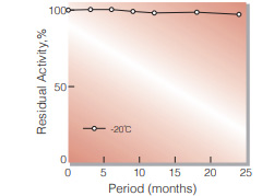
Fig.1. Stability (Powder form)
(kept under dry conditions)
-
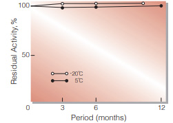
Fig.2. Stability (Powder form)
(kept under dry conditions)
-
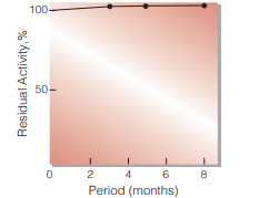
Fig.3. Stability (Liquid form at 5℃)
enzyme concentration:5,000U/ml composition:3.2M ammonium sulfate,pH6.0
-
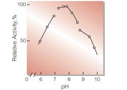
Fig.4. pH-Activity
30℃ in the following buffer solution: pH5.7-6.8, 15mM Veronal-CH3COONaHCI;pH6.8-8.5,50mM Tris-HCI; pH8.5-9.5, 50mM glycine-NaOH
-
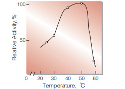
Fig.5. Temperature activity
(in 50mM Tris-HCI buffer, pH7.8)
-
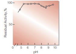
Fig.6. pH-Stability
30℃, 17hr-treatment with the following buffer solution: pH5.0-7.8, 30mM VeronalCH3COONa-HCI;pH7.5-8.5, 0.1M Tris-HCI; pH8.5-9.5,0.1M glycine-NaOH
-
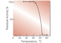
Fig.7. Thermal stability
30min-treatment with 5.0mM glycineNaOH buffer, pH8.0, containing 0.1% of bovine serum albumin
活性測定法(Japanese)
1. 原理
NADHの生成量を340nmの吸光度の変化で測定する。
2.定義
下記条件で1分間に1マイクロモルのNADHを生成する酵素量を1単位(U)とする。
3.試薬
- 55mM Tris-HCl 緩衝液,pH7.8(3.3mMのMgCl2を含む)
- 60mM NAD+水溶液(用時調製)
- 0.1M G-6-P(glucose-6-phosphate)水溶液(用時調製)
酵素溶液:酵素標品を予め氷冷した0.1%牛血清アルブミン(BSA)を含む5mM Tris-HCl緩衝液,pH7.5で溶解し,分析直前に同緩衝液で0.05〜0.20U/mlに希釈する。
4.手順
1.下記反応混液をキュベット(d=1.0cm)に調製し,30℃で約5分間予備加温する。
| 2.7ml | Tris-HCl 緩衝液 | (A) |
| 0.1ml | NAD+水溶液 | (B) |
| 0.1ml | 基質溶液 | (C) |
2.酵素溶液0.1mlを添加し,ゆるやかに混和後,水を対照に30℃に制御された分光光度計で340nmの吸光度変化を4〜5分間記録し,その初期直線部分から1分間当りの吸光度変化を求める(ΔODtest)。
3.盲検は酵素溶液の代りに酵素希釈液(0.1%BSAを含む5mM Tris-HCl緩衝液,pH7.5)を0.1ml加え,上記同様に操作を行って1分間当りの吸光度変化を求める(ΔODblank)。
5.計算式
U/ml =
-
ΔOD/min (ΔOD test−ΔOD blank)×3.0(ml)×希釈倍率
6.22×1.0×0.1(ml)
| = ΔOD/min×4.82×希釈倍率 | |
| U/mg | = U/ml×1/C |
| 6.22 | : NADHのミリモル分子吸光係数(cm2/micromole) |
| 1.0 | : 光路長(cm) |
| C | : 溶解時の酵素濃度(c mg/ml) |
CONTACT
お問い合わせ-
各種製品に関するご質問・ご相談はこちらよりお問い合わせください。