-
GYK-311
GLYCEROL KINASE from Microorganism
PREPARATION and SPECIFICATION
| Appearance | White amorphous powder, lyophilized | |
|---|---|---|
| Activity | GradeⅢ 30U/mg-solid or more | |
| Contaminants | Catalase | ≤1.0×10⁻¹% |
| NADH oxidase | ≤1.0×10⁻³% | |
| Adenosine triphosphatase | ≤1.0×10⁻³% | |
PROPERTIES
| Stability | Stable at −20℃ for at least one year (Fig.1) |
|---|---|
| Molecular weight | approx. 220,000 (by gel filtration) |
| Structure | Four subunits of approx. 58,000 |
| Isoelectric point | 4.3 |
| Michaelis constants | 9.4×10⁻⁵M (Glycerol), 1.3×10⁻⁵M (ATP), 2.1×10⁻³M (Dihydroxyacetone) |
| Inhibitors | p-Chloromercuribenzoate, Hg++, Ag+ |
| Optimum pH | 10.0 (Fig.2) |
| Optimum temperature | 70℃ (Fig.3) |
| pH Stability | pH 5.5−10.0 (25℃, 20hr) (Fig.4) |
| Thermal stability | below 65℃ (pH 7.5, 30min) (Fig.5) |
| Substrate specificity | (Table 1) |
| Effect of various chemicals | (Table 2) |
APPLICATIONS
This enzyme is useful for enzymatic determination of glycerol and triglyceride when coupled with glycerol-3-phosphate oxidase (=G-3-P oxidase, G3O-321) or pyruvate kinase and lactate dehydrogenase (LCD-209, LCD-211, LCD-221), lipoprotein lipase (LPL-311, LPL-314) in clinical analysis.
ASSAY
Principle


The appearance of quinoneimine dye is measured at 500nm by spectrophotometry.
Unit definition
One unit causes the formation of one micromole of hydrogen peroxide (half a micromole of quinoneimine dye) per minute under the conditions described below.
Method
Reagents
| A. Glycerol solution | 0.3M (Should be prepared fresh) | |
|---|---|---|
| B. 4-AA solution | 0.1 % (100mg of 4-aminoantipyrine / 100ml of H2O) | |
| C. Phenol solution | 0.1 % (100mg of phenol / 100ml of H2O) | |
| D. Peroxidase solution | 20mg Peroxidase (110 purpurogallin units/mg)/100ml of H2O | |
| E. G-3-POD solution | 20U/ml (dissolve in 200 mM HEPES buffer, pH 7.9) | |
| F. Buffer solution | 200mM HEPES, pH 7.9 contg. 20mM MgCl2 and 40mM ATP (should be prepared freshly) | |
| G. Enzyme diluent | 20mM K-phosphate buffer, pH 7.5 | |
Procedure
1. Prepare the following working solution in a brownish bottle and store on ice.
| 10ml | 4-AA solution | (B) |
| 20ml | Phenol solution | (C) |
| 20ml | Peroxidase solution | (D) |
| 40ml | G-3-POD solution | (E) |
| 10ml | Buffer solution | (F) |
| Concentration in assay mixture | |
|---|---|
| HEPES buffer | 95.2 mM |
| Glycerol | 4.76 mM |
| ATP | 3.81 mM |
| MgCl2 | 1.90 mM |
| 4-AA | 0.469 mM |
| Phenol | 2.02 mM |
| Peroxidase | ca.5.2 U/ml |
| G-3-POD | ca.7.6 U/ml |
2. Pipette 3.0 ml of working solution in a cuvette (d=1.0cm).
3. Add 0.1ml of enzyme solution*, mix by gently inversion and equilibrate at 37℃ for about 5 minutes.
4. Add 0.05ml of glycerol solution (A) and mix by gentle inversion.
5. Record the optical density at 500nm against water for 3 to 4 minutes in a spectrophotometer thermostated at 37℃, and calculate ΔOD per minute from the initial portion of the curve (ΔOD test).
At the same time, measure the blank rate (ΔOD blank) by the same method as test except the enzyme diluent is added instead of the enzyme solution.
*Dissolve the enzyme preparation in ice-cold enzyme diluent (G) and dilute to 0.2−0.4U/ml with the same buffer, immediately before assay.
Calculation
Activity can be calculated by using the following formula :
Volume activity (U/ml) =
-
ΔOD/min (ΔOD/min (OD test−OD blank)×Vt×df
13.3×1/2×1.0×Vs
= ΔOD/min×4.74×df
Weight activity (U/mg) = (U/ml)×1/C
| Vt | : Total volume (3.15ml) |
| Vs | : Sample volume (0.1ml) |
| 13.3 | : Millimolar extinction coefficient of quinoneimine dye under the assay condition (㎠/micromole) |
| 1/2 | : Factor based o the fact that one mole of H2O2 produces half a mole of quinoneimine dye |
| 1.0 | : Light path length (cm) |
| df | : Dilution factor |
| C | : Enzyme concentration in dissolution (c mg/ml) |
REFERENCES
1) H.-S.Huang, T.Yoshida, Y.Meng, T.Kabashima, K.Ito, Y.Nishiya, Y.Kawamura, and T.Yoshimoto; J.Ferment.Bioeng., 83, 328 (1997).
Table 1. Substrate Specificity of Glycerol kinase
[Pyruvate kinase-Lactate dehydrogenase system with 50mM HEPES buffer, pH 7.9]
-
Substrate (4.5mM) Relative activity(%) Glycerol 100 Glycerol-α-monochlorohydrin 0.1 Ethylene glycol - 1,2-Propanediol - 1,3-Propanediol 0.2 1,3-Butanediol - 1,4-Butanediol 0.1 -
Substrate (4.5mM) Relative activity(%) 2,3-Butanediol 0.2 D-Mannitol - D-Sorbitol - D-Glucose - Ribitol - Methanol - Ethanol -
Table 2. Effect of Various Chemicals on Glycerol kinase
[The enzyme dissolved in 20mM K-phosphate buffer, pH 7.5 (100U/ml) was incubated with each chemical at 25℃ for 1hr.]
-
Chemical Concn.(mM) Residual
activity(%)None - 100 Metal salt MgCl2 1.0 100 CaCl2 102 Ba(OAc)2 101 FeSO4 98 FeCl3 89 CoCl2 104 MnCl2 99 ZnCl2 103 Cd(OAc)2 101 NiCl2 98 CuSO4 99 Pb(OAc)2 100 AgNO3 10 HgCl2 2 Dithiothreitol 1.0 100 PCMB 1.0 0 -
Chemical Concn.(mM) Residual
activity(%)MIA 1.0 101 NaF 1.0 100 NaN3 1.0 106 EDTA 5.0 100 o-Phenanthroline 1.0 102 α,α′-Dipyridyl 1.0 101 Borate 50 103 IAA 1.0 99 NEM 1.0 100 Hydroxylamine 1.0 99 Triton X-100 1.0% 103 Brij 35 0.1% 104 Tween 20 0.1% 103 Span 20 0.1% 102 Na-cholate 0.5% 105 SDS 0.5% 1 DAC 0.5% 84
Ac, CH3CO; PCMB, p-Chloromercuribenzoate; MIA, Monoiodoacetate; EDTA, Ethylenediaminetetraacetate; IAA, Iodoacetamide; NEM, N-Ethylmaleimide; SDS, Sodium dodecyl sulfate; DAC, Dimethylbenzylalkylammonium chloride.
-
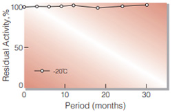
Fig.1. Stability (Powder form)
(kept under dry conditions)
-
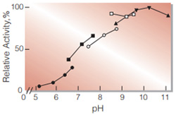
Fig.2. pH-Activity
37℃ 10min-reaction in 45mM buffer solution: pH5.2-6.7,MES:pH6.6-7.7,HEPES; pH7.5-8.7,TAPS;pH8.5-9.6,CHES; pH8.7-11.2,Glycine-NaOH
-
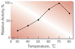
Fig.3. Temperature activity
(10min-reaction in 45mM HEPES buffer,pH7.9)
-
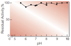
Fig.4. pH-Stability
enzyme concn. ca.300U/ml 25℃ 20hr-treatment in 50mM buffer solution: pH5.6-7.1,MES;pH7.1-8.0,HEPES;pH8.0-9.0, TAPS;pH9.0-10.0,CHES
-
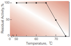
Fig.5. Thermalstability
enzyme concn. ca.300U/ml 30min-treatment with 20mM K-phosphate buffer, pH7.5
活性測定法(Japanese)
1. 原理
4-AminoantipyrineとPhenolの酸化縮合生成物であるQuinoneimine色素を500 nmで測定し, 上記反応で生成したH2O2量を定量する。
2.定義
下記条件下で1分間に1マイクロモルのH2O2を生成する酵素量を1単位(U)とする。
3.試薬
- 0.3M グリセロール溶液(用時調製)
- 4-AA溶液, 0.1 %(100mgの4-アミノアンチピリンを100mlの蒸留水で溶解する。)
- フェノール溶液,0.1%(100mgのフェノールを100mlの蒸留水で溶解する。)
- ペルオキシダーゼ溶液[25mgのペルオキシダーゼ(110プルプロガリン単位/mg)を約100mlの蒸留水で溶解する。]
- G-3-POD溶液, 20U/ml(200mM HEPES緩衝液,pH7.9で溶解する。)
- 緩衝液(20 mM MgCl2と40mM ATPを含む200mM HEPES緩衝液, pH 7.9)(用時調製)
酵素溶液:酵素標品を予め氷冷した20mM Kリン酸緩衝液, pH7.5で分析直前に0.2〜0.4 U/mlに希釈する。
4.手順
1.下記反応混液を褐色ビンに調製する(褐色ビンにて氷冷保存)。
| 10ml | 4-AA溶液 | (B) |
| 20ml | フェノール溶液 | (C) |
| 20ml | ペルオキシダーゼ溶液 | (D) |
| 40ml | G-3-POD溶液 | (E) |
| 10ml | 緩衝液 | (F) |
2.上記反応混液をキュベット(d=1cm)に3.0ml採り, 酵素液0.1mlを加え, ゆるやかに混和後, 37℃で約5分間予備加温する。
3.グリセロール溶液(A)0.05mlを添加し, ゆるやかに混和後, 水を対照に37℃に制御された分光光度計で500nmの吸光度変化を3〜4分間記録し, その初期直線部分から1分間当たりの吸光度変化を求める(ΔODtest)。
4.盲検は反応混液①に酵素溶液の代わりに酵素希釈液(20mM Kリン酸緩衝液、pH7.5)を0.1ml加え, 上記同様に操作を行って,1分間当たりの吸光度変化を求める(ΔODblank)。
5.計算式
U/ml =
-
ΔOD/min (OD test−OD blank)×3.15(ml)×希釈倍率
13.3×1/2×1.0×0.1(ml)
| = ΔOD/min×4.74×希釈倍率 | |
| U/mg | = U/ml×1/C |
| 13.3 | : Quinoneimine色素の上記測定条件下でのミリモル分子吸光係数(cm2/micromole) |
| 1/2 | : 酵素反応で生成したH2O21分子から形成するQuinoneimine色素は1/2分子である事による係数 |
| 1.0 | : 光路長(cm) |
| C | : 溶解時の酵素濃度(c mg/ml) |
CONTACT
お問い合わせ-
各種製品に関するご質問・ご相談はこちらよりお問い合わせください。