PPC-301
PHOSPHOENOLPYRUVATE CARBOXYLASE from Microorganism
PREPARATION and SPECIFICATION
| Appearance | White amorphous powder, lyophilized | |
|---|---|---|
| Activity | GradeⅢ 5.0U/mg-solid or more | |
| Contaminants | Lactate dehydrogenase | ≤1.0×10⁻³% |
| Pyruvate kinase | ≤0.05% | |
| Stabilizers | BSA, sugar alcohols | |
PROPERTIES
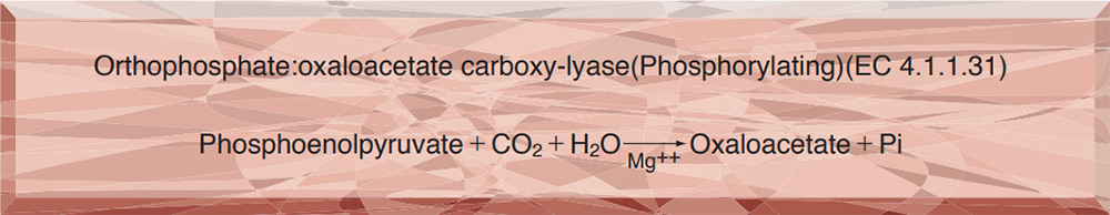
| Stability | Stable at −20℃ for at least one year (Fig.1) |
|---|---|
| Molecular weight | approx. 390,000 (by gel filtration) |
| Isoelectric point | 6.0±0.1 |
| Structure | 4 Subunits (M.W.100,000) per enzyme molecule |
| Michaelis constant | 1.9×10⁻⁴M (Phosphoenolpyruvate) |
| Optimum pH | 7.5−8.0 (Fig.2) |
| Optimum temperature | 60℃ (Fig.3) |
| pH Stability | pH 5.0−8.0 (25℃, 24hr) (Fig.4) |
| Thermal stability | below 40℃ (pH 7.0, 15min) (Fig.5) |
APPLICATIONS
This enzyme is useful for enzymatic determination of carbon dioxide when coupled with malate dehydrogenase (MAD-211) in clinical analysis.
ASSAY
Principle

The disappearance of NADH is measured at 340nm by spectrophotometry.
Unit definition
One unit causes the oxidation of one micromole of NADH per minute under the conditions described below.
Method
Reagents
| A. Buffer solution | 0.1M Tris-HCl Buffer, pH 8.0 |
|---|---|
| B. Na2CO3 solution | 0.1M[Dissolve 1.06g of Na22CO3(MW=105.99)/100ml of H2O] |
| C. K-Phosphoenolpyruvate solution | 32mM[Dissolve 33.0mg of PEP・K(MW=206.1)/5ml of H2O](Should be prepared fresh) |
| D. MgSO4 solution | 1M[Dissolve 4.93g of MgSO4・7H2O(MW=246.48)/20 ml of H2O] |
| E. NADH solution | 1.4mM[Dissolve 5.34mg of NADH・3H2O(MW=763)/5ml of H2O] |
| F. MDH solution | ca.100U/ml[Dissolve malate dehydrogenase (TOYOBO GradeⅡ) to approx.100U/ml with 20mM Tris-HCl Buffer,pH 8.0](Should be prepared fresh) |
| G. Enzyme diluent | 20mM K-phosphate buffer, pH 7.0 |
Procedure
1.Prepare the following reaction mixture in a cuvette (d=1.0cm) and equilibrate at 30℃ for about 5 minutes.
| 1.77ml | Buffer solution | (A) |
|---|---|---|
| 0.3 ml | Na2CO3 solution | (B) |
| 0.3 ml | K-Phosphoenolpyruvate solution | (C) |
| 0.03ml | MgSO4 solution | (D) |
| 0.3 ml | NADH solution | (E) |
| 0.3 ml | MDH solution | (F) |
| Concentration in assay mixture | |
|---|---|
| K-Phosphoenolpyruvate | 3.1 mM |
| Tris-HCl | 57 mM |
| Na2CO3 | 9.7 mM |
| MgSO4 | 9.7 mM |
| NADH | 0.14mM |
| MDH | 9.7 U/ml |
| K-Phosphate | 0.65mM |
2.Add 0.1ml of the enzyme solution* and mix by gentle inversion.
3.Record the decrease in optical density at 340nm against water for 3 to 4 minutes in a spectrophotometer thermostated at 30℃, and calculate the ΔOD per minute from initial liner portion of the curve (ΔOD test).
At the same time, measure the blank rate (ΔOD blank) by using the same method as the test except that the enzyme diluent (G) is added instead of the enzyme solution.
*Dissolve the enzyme preparation in ice-cold enzyme diluent (G) and dilute to 0.2−0.7U/ml with the same buffer and store on ice.
Calculation
Activity can be calculated by using the following formula :
Volume activity (U/ml) =
-
ΔOD/min (ΔOD test−ΔOD blank)×Vt×df
6.22×1.0×Vs
= ΔOD/min×4.98×df
Weight activity (U/mg) = (U/ml)×1/C
| Vt | : Total volume (3.1ml) |
| Vs | : Sample volume (0.1ml) |
| 6.22 | : Millimolar extinction coefficient of NADH (cm2/micromole) |
| 1.0 | : Light path length (cm) |
| df | : Dilution factor |
| C | : Enzyme concentration in dissolution (c mg/ml) |
REFERENCES
1) W.Wilson, P.Jesyk, R.Rand and R.D.Bevill; Clin.Chem.,19, 640(1973)
2) R.L.Forrester, L.J.Wataji, D.A.Silverman and K.J.Pierre; Clin.Chem.,22, 243(1976)
Table 1. Effect of Various Chemicals on Phosphoenolpyruvate carboxylase
[The enzyme solution dissolved in 20mM K-phosphate buffer, pH 7.0 (20U/ml) was incubated with each chemical at 25℃ for 1hr.]
-
Chemical Concn.(mM) Residual
activity(%)None - 100 Metal salt 2.0 MgCl2 105 CaCl2 105 Ba(OAc)2 103 FeCl3 92 CoCl2 106 MnCl2 107 ZnSO4 103 Cd(OAc)2 104 NiCl2 0 CuSO4 0 Pb(OAc)2 105 AgNO3 0 HgCl2 0 MIA 2.0 60 2-Mercaptoethanol 2.0 101 -
Chemical Concn.(mM) Residual
activity(%)PCMB 0.1 80 NEM 2.0 87 IAA 2.0 90 Hydroxylamine 2.0 95 EDTA 5.0 100 o-Phenanthroline 2.0 103 α,α′-Dipyridyl 2.0 109 Borate 5.0 103 NaF 2.0 106 NaN3 2.0 106 Triton X-100 0.10% 111 Brij 35 0.10% 110 Tween 20 0.10% 112 Span 20 0.10% 109 Na-cholate 0.10% 108 SDS 0.05% 1 DAC 0.05% 99
Ac, CH3CO; PCMB, p-Chloromercuribenzoate; MIA, Monoiodoacetate; EDTA, Ethylenediaminetetraacetate; IAA, Iodoacetamide; NEM, N-Ethylmaleimide; SDS, Sodium dodecyl sulfate; DAC, Dimethylbenzylallkylammonium chloride.
-
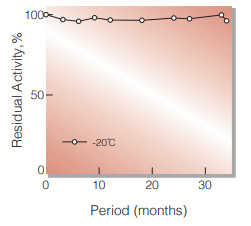
Fig.1. Stability (Powder form)
(kept under dry conditions)
-
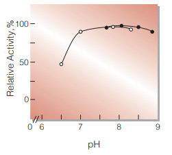
Fig.2. pH-Activity
30℃, in 50mM buffer solution: pH6.0-8.5, MES: pH7.5-9.0, Tris-HCI
-
Fig.3. Temperature activity
(in 20mM K-phosphate buffer,pH7.0)
-
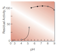
Fig.4. pH-Stability
25℃,24hr-treatment with 50mM buffer solution contg. 10mM MgSO4: pH3.0-5.0, Acetate;pH5.0-8.0, K-phosphate; pH8.0-9.0, Tris-HCI
-
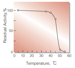
Fig.5. Thermal stability
15min-treatment with 20mM K-phosphate buffer,pH7.0 enzyme concn.: 2.0U/ml
活性測定法 (Japanese)
1. 原理
NADHの減少量を340nmにおける吸光度の変化で測定する。
2.定義
下記条件で1分間に1マイクロモルのNADHを酸化する酵素量を1単位(U)とする。
3.試薬
- 0.1M Tris-HCl緩衝液,pH8.0
- 0.1M Na2CO3水溶液(1.06gの無水炭酸ナトリウム(MW=105.99)を蒸留水100mlに溶解する。)
- 32.0mM K-Phosphoenolpyruvate水溶液(33.0mgのK-phosphoenolpyruvate(MW=206.1)を蒸留水5.0mlに溶解する。)(用時調製)
- 1.0M MgSO4水溶液(4.93gのMgSO4・7H2O(MW=246.48)を蒸留水20mlに溶解する。)
- 1.4mM NADH水溶液(5.34mgのNADH・Na2(MW=763)を蒸留水5.0mlに溶解する。)
- 100U/ml Malate dehydrogenase溶液(リンゴ酸脱水素酵素(東洋紡製 GradeⅡ)を20mM TrisHCl pH8.0で溶解する。)(用時調製)
酵素溶液:酵素標品を予め氷冷した20mM K-リン酸緩衝液,pH7.0で溶解し,同緩衝液で0.2〜0.7U/mlに希釈する。
4.手順
1.下記反応混液をキュベット(d=1.0cm)に調製し,30℃で約5分間予備加温する。
| 1.77 | Tris-HCl緩衝液 | (A) |
| 0.30 | Na2CO3 水溶液 | (B) |
| 0.30 | K-Phosphoenolpyruvate水溶液 | (C) |
| 0.03 | MgSO4水溶液 | (D) |
| 0.30 | NADH水溶液 | (E) |
| 0.30 | Malate dehydrogenase溶液 | (F) |
2.酵素溶液0.1mlを添加し,ゆるやかに混和後,水を対照に30℃に制御された分光光度計で340nmの吸光度変化を3〜4分間記録し,その初期直線部分から1分間当りの吸光度変化を求める(ΔODtest)。
3.盲検は,酵素溶液の代わりに酵素希釈液を0.1ml加え,上記同様に操作を行って1分間当たりの吸光度変化を求める(ΔODblank)。
5.計算式
U/ml =
-
ΔOD/min (ΔOD test−ΔOD blank)×3.1(ml)×希釈倍率
6.22×1.0×0.1(ml)
| = ΔOD/min×4.984×希釈倍率 | |
| U/mg | = U/ml×1/C |
| 6.22 | : NADHのミリモル分子吸光係数(cm2/micromole) |
| 1.0 | : 光路長(cm) |
| C | : 溶解時の酵素濃度(c mg/ml) |
CONTACT
お問い合わせ-
各種製品に関するご質問・ご相談はこちらよりお問い合わせください。