PYO-311
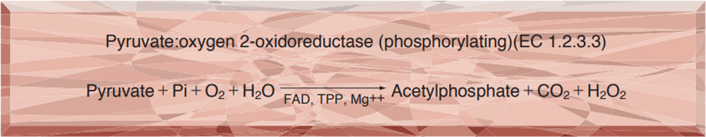
PYRUVATE OXIDASE from Microorganism
PREPARATION and SPECIFICATION
| Appearance | Yellowish amorphous powder, lyophilized | |
|---|---|---|
| Activity | GradeⅢ 1.5U/mg-solid or more | |
| Contaminants | ATPase | ≤5.0×10⁻²% |
| GOT, GPT | ≤5.0×10⁻²% | |
| Stabilizers | Sugars, FAD | |
PROPERTIES
| Stability | Stable at −20℃ for at least one year (Fig.1) |
|---|---|
| Molecular weight | approx. 260,000 |
| Isoelectric point | 4.3 |
| Michaelis constant | 3.4×10⁻⁴ M (Pyruvate) |
| Inhibitors | Fe++,Zn++,Cu++,Ag+,Hg++ |
| Optimum pH | 5.7 (Fig.2) |
| Optimum temperature | 65℃ (Fig.3) |
| pH Stability | pH 5.7−6.5 (25℃, 20hr) (Fig.4) |
| Thermal stability | below 45℃ (pH 6.0, 15min) (Fig.5) |
| Substrate specificity | (Table 1) |
| Effect of various chemicals | (Table 2) |
APPLICATIONS
This enzyme is useful for enzymatic determination of pyruvate, GOT, GPT in clinical analysis.
ASSAY
Principle
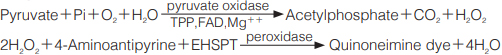
The appearance of quinoneimine dye is measured at 550nm by spectrophotometry.
Unit definition
One unit causes the formation of one micromole of hydrogen peroxide (half a micromole of quinoneimine dye) per minute under the conditions described below.
Method
Reagents
| A. Pyruvate solution | 0.3M[378mg of Pyruvate・K salt (MW=126.15)/10ml of H2O] |
|---|---|
| B. K-phosphate buffer, pH 5.9 | 0.15M |
| C. 4-Aminoantipyrine solution | 0.15%(150mg of 4-Aminoantipyrine/100ml of H2O) |
| D. EHSPT (TOOS) solution | 0.3%[300mg of EHSPT (N-Ethyl-N-(2-hydroxy-3-sulfopropyl)-m-toluidine)/100ml of H2O] |
| E. TPP solution | 3mM[13.8mg of TPP (Thiamine pyrophosphate)(MW=460.77)/10ml of H2O] |
| F. FAD solution | 0.15mM[1.3mg of FAD・2Na salt (MW=865.55)/10ml of H2O] |
| G. EDTA solution | 15mM[590mg of EDTA・2Na salt (MW=394.22)/100ml of H2O] |
| H. MgSO4 solution | 0.15M[3.4g of MgSO4・7H2O(246.48)/100ml of H2O] |
| I. Peroxidase solution | 50U/ml[45mg of peroxidase (110purpurogallin units/mg)/100ml of H2O] |
| J. Enzyme diluent | 50mM K-phosphate buffer, pH 5.7 |
Procedure
1.Prepare the following working solution in a brownish bottle and store on ice.
| 10ml | K-phosphate buffer, pH 5.9 | (B) |
|---|---|---|
| 2ml | 4-Aminoantipyrine solution | (C) |
| 2ml | EHSPT solution | (D) |
| 2ml | TPP solutionn | (E) |
| 2ml | FAD solution | (F) |
| 2ml | EDTA solution | (G) |
| 2ml | MgSO4 solution | (H) |
| 3ml | Peroxidase | (I) |
| Concentration in assay mixture | |
|---|---|
| Pyruvate | 48 mM |
| K-phosphate buffer | 50 mM |
| 4-Aminoantipyrine | 0.48mM |
| EHSPT | 0.58mM |
| TPP | 0.19mM |
| FAD | 0.01mM |
| EDTA | 0.97mM |
| MgSO4 | 9.7 mM |
| Peroxidase | ca.4.8 U/ml |
2.Pipette 2.5ml of working solution into a cuvette (d=1.0cm), add 0.5ml of pyruvate solution (A), and equilibrate at 37℃ for about 5minutes.
3.Add 0.1ml of the enzyme solution* and mix by gentle inversion.
4.Record the increase in optical density at 550nm against water for 3 to 4 minutes in a spectrophotometer thermostated at 37℃, and calculate the ΔOD per minute from the initial linear portion of the curve (ΔOD test).
At the same time, measure the blank rate (ΔOD blank) by using the same method as the test except that the enzyme diluent is added instead of the enzyme solution.
*Dissolve the enzyme preparation in ice-cold enzyme diluent (J), dilute to 0.1−0.5U/ml with the same buffer and store on ice.
Calculation
Activity can be calculated by using the following formula :
Volume activity (U/ml) =
-
ΔOD/min (ΔOD test−ΔOD blank)×Vt×df
36.88×1/2×1.0×Vs
= ΔOD/min×1.68×df
Weight activity (U/mg) = (U/ml)×1/C
| Vt | : Total volume (3.10ml) |
| Vs | : Sample volume (0.10ml) |
| 36.88 | : Millimolar extinction coefficient of quinoneimine dye under the assay condition (cm2/micromole) |
| 1/2 | : Factor based on the fact that one mole of H2O2 produces half a mole of quinoneimine dye. |
| 1.0 | : Light path length (cm) |
| df | : Dilution factor |
| C | : Enzyme concentration in dissolution (c mg/ml) |
REFERENCES
1) L.P.Hager, D.M.Geller and F.Lipman; Fed.Proc.,13, 734 (1954).
2) B.Sedewitz, K.H.Schleifer and F.Gotz; J.Bacteriol,160, 273 (1984).
3) B.Sedewitz, K.H.Schleifer and F.Gotz; J.Bacteriol,160, 462 (1984).
Table 1. Substrate Specificity of Pyruvate oxidase
-
Substrate(50mM) Relative activity(%) Pyruvate 100 α-Ketobutyrate 5.8 α-Ketoglutarate 0 Oxaloacetate 0 DL-Lactate 0 -
Substrate(50mM) Relative activity(%) Acetate 0 Acetoacetate 0 L-Alanine 0 L-Aspartate 0
Table 2. Effect of Various Chemicals on Pyruvate oxidase
[The enzyme dissolved in 50mM K-phosphate, pH 6.0 (10U/ml) was incubated with each chemical at 25℃ for 1hr]
-
Chemical Concn.(mM) Residual
activity(%)None - 100 Metal salt 2.0 MgCl2 96 CaCl2 93 Ba(OAc)2 97 FeCl3 8.4 CoCl2 84 MnCl2 76 ZnSO4 48 Cd(OAc)2 86 NiCl2 119 CuSO4 0.9 Pb(OAc)2 33 AgNO3 0 HgCl2 0 PCMB 1.0 66 MIA 2.0 96 -
Chemical Concn.(mM) Residual
activity(%)NaF 2.0 100 NaN3 20 94 EDTA 5.0 107 o-Phenanthroline 2.0 97 α,α′-Dipyridyl 1.0 95 Borate 50 102 IAA 2.0 102 NEM 2.0 104 Hydroxylamin 2.0 98 Triton X-100 0.10% 143 Brij 35 0.10% 133 Tween 20 0.10% 146 Span 20 0.10% 121 Na-cholate 0.10% 116 SDS 0.05% 85 DAC 0.05% 53
Ac, CH3CO; PCMB, p-Chloromercuribenzoate; MIA, Monoiodoacetate; EDTA, Ethylenediaminetetraacetate;
IAA, Iodoacetamide; NEM, N-Ethylmaleimide; SDS, Sodium dodecyl sulfate; DAC, Dimethylbenzylallkylammonium chloride.
-
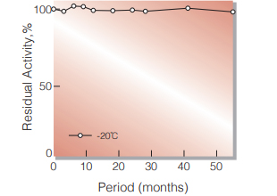
Fig.1. Stability (Power form)
(kept under dry conditions)
-
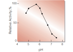
Fig.2. pH-Activity
(37℃ in 50mM K-phosphate buffer)
-
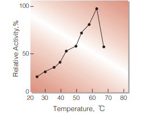
Fig.3. Temperature activity
(in 50mM K-phosphate buffer, pH5.7)
-
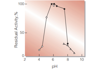
Fig.4. pH-Stability
25℃,20hr-treatment with 50mM buffer solution (contg. 10mM MgSO4. 10μM FAD, 0.2mM TPP):pH4.0-6.0, acetate;pH5.7-8.0 K-phosphate; pH7.5-9.0,Tris-HCI
-
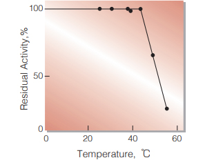
Fig.5. Thermal stability
15min-treatment with 50mM K-phosphate buffer(cong. 10mM MgSO4. 10μM FAD, 0.2mM TPP), pH6.0. enzyme concn.:10U/ml
活性測定法(Japanese)
1. 原理
4-AminoantipyrineとEHSPTの酸化縮合生成物である Quinoneimine色素を550nmで測定し,上記反応で生成したH2O2量を定量する。
2.定義
下記条件で1分間に1マイクロモルのH2O2を生成する酵素量を1単位(U)とする。
3.試薬
- 0.3Mピルビン酸水溶液〔378mgのピルビン酸・K塩 (MW=126.15)を10mlの蒸留水に溶解する。〕
- 0.15Mリン酸カリウム緩衝液,pH5.9
- 0.15%4-AA水溶液(150mgの4-アミノアンチピリン を100mlの蒸留水に溶解する。)
- 0.3%EHSPT(TOOS)水溶液〔300mgのEHSPT を100mlの蒸留水に溶解する〕
- 3.0mM TPP水溶液〔13.8mgのTPP(MW= 460.77)を10mlの蒸留水に溶解する。〕
- 0.15mM FAD水溶液〔1.3mgのFAD・2Na塩 (MW=865.55)を10mlの蒸留水に溶解する。〕
- 15mM EDTA水溶液〔590mgのEDTA・2Na塩 (MW=394.22)を100mlの蒸留水に溶解する。〕
- 0.15M MgSO4水溶液〔3.4gのMgSO4・7H2O (MW=246.48)を100mlの蒸留水に溶解する。〕
- 50U/ml POD水溶液〔45mgペルオキシダーゼ (POD)(110プルプロガリン単位/mg)を100mlの蒸留水に溶解する。〕
酵素溶液:酵素標品を予め氷冷した50mM K-リン酸緩衝液,pH5.7で溶解し,分析直前に同緩衝液で0.1〜0.5U/mlに希釈する。
4.手順
1.下記反応混液を調製する(褐色瓶にて氷冷保存)。
| 10ml | K-リン酸緩衝液 | (B) |
| 2ml | 4-AA水溶液 | (C) |
| 2ml | EHSPT水溶液 | (D) |
| 2ml | TPP水溶液 | (E) |
| 2ml | FAD水溶液 | (F) |
| 2ml | EDTA水溶液 | (G) |
| 2ml | MgSO4水溶液 | (H) |
| 3ml | POD水溶液 | (I) |
2.反応混液2.5mlをキュベット(d=1.0cm)に採り,ピルビン酸水溶液(A)0.5mlを添加し,37℃で約5分間予備加温する。
3.酵素溶液0.1mlを添加し,ゆるやかに混和後,水を対照に37℃に制御された分光光度計で550nmの吸光度 変化を3~4分間記録し,その初期直線部分から1分間 当りの吸光度変化を求める(ΔODtest)。
4.盲検は反応混液①に酵素溶液の代わりに酵素希釈液(50mM K-リン酸緩衝液,pH5.7)を0.1ml加え,上記同様に操作を行って,1分間当たりの吸光度変化を求める(ΔODblank)。
5.計算式
U/ml =
-
ΔOD/min (ΔOD test−ΔOD blank)×3.1(ml)×希釈倍率
36.88×1/2×1.0×0.1(ml)
| = ΔOD/min×1.68×希釈倍率 | |
| U/mg | = U/ml×1/C |
| 36.88 | : Quinoneimine色素の上記測定条件下でのミリモル分子吸光係数(cm2/micromole) |
| 1/2 | : 酵素反応で生成したH2O2の1分子から形成するQuinoneimine色素は1/2分子である事による係数。 |
| 1.0 | : 光路長(cm) |
| C | : 溶解時の酵素濃度(c mg/ml) |
CONTACT
お問い合わせ-
各種製品に関するご質問・ご相談はこちらよりお問い合わせください。