-
BGH-101
β-GLUCOSIDASE from Sweet almond

PREPARATION and SPECIFICATION
| Appearance | Light yellow amorphous powder, lyophilized | |
|---|---|---|
| Activity | GradeⅠ 15U/mg-solid or more (containing approx. 30% of BSA) |
|
| Contaminant | α-Amylase | ≤5.0×10⁻⁴% |
| Stabilizers | BSA, glutathione (reduced) | |
PROPERTIES
| Stability | Stable at −20℃ for at least one year(Fig.1) |
|---|---|
| Molecular weight | approx. 110,000 |
| Isoelectric point | 7.3 1) |
| Michaelis constants | 2.8×10⁻³M (p-Nitrophenyl-β-D-glucopyranoside), 3.3×10⁻³M (2,4-Dichlorophenyl-β-D-glucopyranoside) |
| Structure | 2 subunits per enzyme molecule |
| Optimum pH | 5.5(Fig.4) |
| Optimum temperature | 50−55℃(Fig.5) |
| pH Stability | pH 6.0−9.0 (25℃, 64hr)(Fig.6) |
| Thermal stability | below 50℃ (pH 7.3, 1hr)(Fig.7) |
| Effect of various chemicals | (Table 1) |
APPLICATIONS
This enzyme is useful for structural investigations of carbohydrates and for the enzymatic determination of α-amylase when coupled with α-glucosidase (AGH-211) in clinical analysis.
ASSAY
Principle

The appearance of p-nitrophenol is measured at 400nm by spectrophotometry.
Unit definition
One unit causes the formation of one micromole of PNP per minute under the conditions described below.
Method
Reagents
| A. Acetate buffer, pH 5.0 (at 25℃) | 0.1M | |
|---|---|---|
| B. PNPG solution | 20mM (603mg p-nitrophenyl-β-D-glucopyranoside/100ml of H2O)(Stable for two weeks if stored at 0−5℃) | |
| C. Na2CO3 solution | 0.2M (21.2g Na2CO3 /1,000ml of H2O) | |
| D. Enzyme diluent | 10mM phosphate buffer, pH 7.0 containing 0.2% of BSA. | |
Procedure
1. Prepare the following reaction mixture in a test tube and equilibrate at 37℃ for about 5 minutes.
| 1.0ml | 0.1M Acetate buffer, pH 5.0 | (A) |
| 0.5ml | Substrate solution | (B) |
| Concentration in assay mixture | |
|---|---|
| Acetate buffer | 50 mM |
| PNPG | 5.0 mM |
| BSA | 0.05mg/ml |
2. Add 0.5ml of the enzyme solution* and mix.
3. After exactly 15 minutes at 37℃, add 2.0ml of Na2CO3 solution (C) to stop the reaction and measure the optical density at 400nm against water (OD test).
At the same time, prepare the blank by first mixing the reaction mixture with 2.0ml of Na2CO3 solution (C) after 15 min-incubation at 37℃, followed by the addition of the enzyme solution (OD blank).
*Dissolve the enzyme preparation in ice-cold 50mM Tris-HCl buffer pH 7.8 (ca. 1mg/ml) and dilute to 0.006−0.022U/ml with the enzyme diluent (D), immediately before assay.
Calculation
Activity can be calculated by using the following formula :
Volume activity (U/ml) =
-
ΔOD (OD test−OD blank)×Vt×df
18.1×1.0×t×Vs
= ΔOD×0.0295×df
Weight activity (U/mg) = (U/ml)×1/C
| Vt | : Total volume (4.0ml) |
| Vs | : Sample volume (0.5ml) |
| 18.1 | : Millimolar extinction coefficient of p-nitrophenol under the assay condition (cm2/micromole) |
| 1.0 | : Light path length (cm) |
| t | : Reaction time (15 minutes) |
| df | : Dilution factor |
| C | : Enzyme concentration in dissolution (c mg/ml) |
REFERENCES
1) A.K.Grover, D.D.Macmurchie and R.J.Cushley; Biochim.Biophys.Acta, 482, 98 (1977).
(Characteristics ofβ-Glucosidase from almond)
2) R.Heyworth and P.G.Walker; Biochem.J., 83, 331 (1962).
3) J.H.Hash and K.W.King; J.Biol.Chem., 232, 395 (1958)
Table 1. Effect of Various Chemicals on β-Glucosidase
[Residual activity after 1 hr-treatment at 30℃.]
-
Chemical Concn.(mM) Residual
activity(%)None - 100 Metal salt 0.5 CaCl2 92.7 FeSO4 94.1 CoCl2 95.5 ZnCl2 95.0 CuSO4 94.5 HgCl2 99.8 CrCl2 93.9 MgSO4 96.8 SnCl2 93.6 CdCl2 93.0 AgNO3 92.7 NiCl2 95.5 -
Chemical Concn.(mM) Residual
activity(%)MnCl2 94.3 BaCl2 93.9 FeCl3 99.8 o-Phenanthroline 0.5 94.3 α,α′-Dipyridyl 0.5 94.3 Borate 25 94.1 PCMB 0.05 94.5 MIA 0.5 89.3 NaF 0.5 96.6 NaN3 10 98.9 EDTA 5.0 96.1 Triton X-100 0.5% 102.3 Na-cholate 0.5% 99.5
PCMB, p-Chloromercuribenzoate; MIA, Monoiodoacetate; EDTA, Ethylenediaminetetraacetate.
-
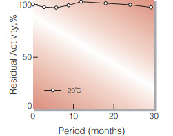
Fig.1. Stability (Powder form)
(kept under dry conditions)
-
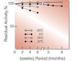
Fig.2. Stability (Powder form)
(kept under dry conditions)
-
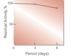
Fig.3. Stability (Liquid form at 25℃)
(enzyme concentration: 1.0mg/ml buffer composition: 50mM Tris-HCI buffer, pH7.8)
-
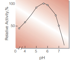
Fig.4. pH-Activity
(37℃.15 min-reaction in 50mM acetate buffer.)
-
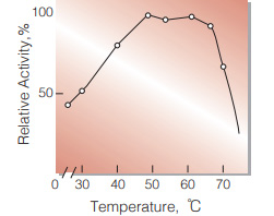
Fig.5.Temperature activity
(15 min-reaction in 50mM acetate buffer, pH5.0)
-
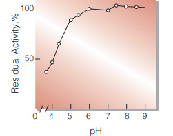
Fig.6. pH-Stability
(25℃, 64hr-treatment with 50mM buffer solution:pH3.5-6.0, acetate; pH6.5-9.0, Tris-HCI)
-
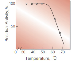
Fig.7. Thermal stability
(1hr-treatment with 50mM Tris-HCI buffer,pH7.3.)
活性測定法(Japanese)
1. 原理
p-Nitrophenolの生成量を400nmの吸光度の変化で 測定する。
2.定義
下記条件下で1分間に1マイクロモルのp-Nitrophenolを生成する酵素量を1単位(U)とする。
3.試薬
- 0.1M酢酸緩衝液, pH5.0(25℃)
- 20mM PNPG水溶液(603mgのP-ニトロフェニルβ-D-グルコピラノシドを100mlの蒸留水に攪拌溶解する)(0〜5℃保存で2週間は使用可能)
- 0.2M Na2CO3溶液(21.2gの無水炭酸ナトリウムを蒸留水に溶解し1,000mlとする)
酵素溶液:酵素標品を予め氷冷した50mM Tris-HCl緩衝液pH7.8で約1mg/mlに溶解し,分析直前に0.2%牛血清アルブミン(BSA)を含む10mMリン酸緩衝液, pH7.0で0.006〜0.022U/mlに希釈する。
4.手順
1.試験管に下記反応混液を調製し,37℃で約5分間予備加温する。
| 1.0ml | 0.1M酢酸緩衝液, pH5.0 | (A) |
| 0.5ml | 基質溶液 | (B) |
2.酵素溶液を0.5mlを加え,反応を開始する。
3.37℃で正確に15分間反応させた後, Na2CO3溶液(C)2.0ml加えて反応を停止させる。この液につき400nmにおける吸光度を測定する(OD test)。
4.盲検は反応混液①を37℃で15分間放置後,Na2CO3溶液(C) 2.0mlを加えて混和し,次いで酵素溶液0.5mlを加えて調製する。以下同様に吸光度を測定する(ODblank)。
5.計算式
U/ml =
-
ΔOD (OD test−OD blank)×4.0(ml)×希釈倍率
18.1×1.0×15(分)×0.5(ml)
| = ΔOD×0.0295×希釈倍率 | |
| U/mg | = U/ml×1/C |
| 18.1 | : p-Nitrophenolの上記測定条件下でのミリモル分子吸光係数(cm2/micromole) |
| 1.0 | : 光路長(cm) |
| C | : 溶解時の酵素濃度(c mg/ml) |
CONTACT
お問い合わせ-
各種製品に関するご質問・ご相談はこちらよりお問い合わせください。