DAD-301
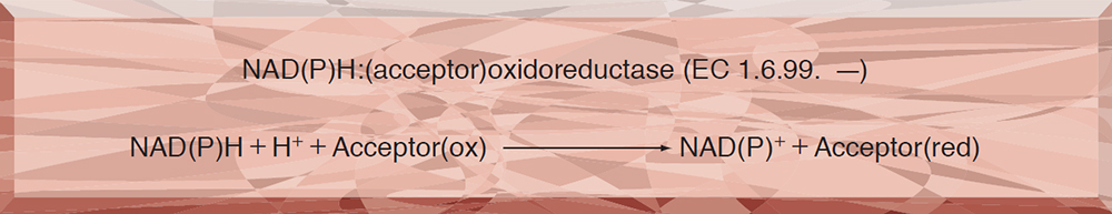
DIAPHORASE from Clostridium sp.
PREPARATION and SPECIFICATION
| Appearance | Yellowish amorphous powder, lyophilized | |
|---|---|---|
| Activity | GradeⅢ 30U/mg-solid or more (containing approx. 15% of stabilizers) |
|
| Contaminants | Myokinase | ≤5.0×10⁻¹% |
| NAD(P)H oxidase | ≤5.0×10⁻¹% | |
| Stabilizers | FMN, NAD(P)H | |
PROPERTIES
| Stability | Stable at −20℃ for at least one year(Fig.1) |
|---|---|
| Molecular weight | 24,000 1) |
| Michaelis constants | 2.0×10⁻⁵M(NADH),6.0×10-6M(NADPH) |
| Structure | One mol of FMN per mol of enzyme 1) |
| Inhibitor | N-Ethylmaleimide |
| Optimum pH | 8.5(Fig.3) |
| Optimum temperature | 50℃(Fig.4) |
| pH Stability | pH7.5(30℃, 3hr)(Fig.5) |
| Thermal stability | below 30℃(pH 7.5, 30min)(Fig.6) |
| Substrate specificty | Either NADH or NADPH can be used as a reductant. The catalytic ratio (NADPH/NADH) is 0.6 in the assay method. Neither oxygen nor cytochrome C can be utilized as a hydrogen acceptor. |
APPLICATIONS
This enzyme is useful for colorimetric determination of NAD(P)H and many dehydrogenases when coupled with various dyes which act as hydrogen acceptors from NAD(P)H.
ASSAY
Principle

The reduction of DCPIP(2,6-dichlorophenol-indophenol) is measured at 600 nm by spectrophotometry.
Unit definition
One unit causes the decrease of one unit absorbance (1.0) of DCPIP per minute under the condeitions described below.
Method
Reagents
| A. Buffer solution | 0.2M Tris-HCl, pH 7.5 |
|---|---|
| B. NADH solution | 6.0mM (Prepare freshly and store on ice) |
| C. DCPIP solution | 1.2mM[3.9mg DCPIP・2H2O/10ml of H2O](Should be prepared fresh) |
| D. Enzyme diluent | Buffer solution(A) containing 0.1% of bovine serum albumin. |
Procedure
1.Prepare the following reaction mixture in a cuvette (d=1.0cm) and equilibrate at 25℃ for about 5 minutes.
| 2.4ml | H2O | |
|---|---|---|
| 0.3ml | Buffer solution | (A) |
| 0.1ml | NADH solution | (B) |
| Concentration in assay mixture | |
|---|---|
| Tris buffer | 27 mM |
| NADH | 0.20 mM |
| DCPIP | 40 μM |
| BSA | ca.33μg/ml |
2.Add 0.1ml each of the enzyme solution* and DCPIP solution (C) in this order and mix by rapid inversion.
3.Record the decrease of optical density at 600nm against water for 2 to 3 minutes in a spectrophotometer thermostated at 25℃, and calculate theΔOD per minute from the initial linear portion of the curve (ΔOD test).
At the same time, measure the blank rate (ΔOD blank) by using the same method as the test except that the enzyme diluent is added instead of the enzyme solution.
*Dissolve the enzyme preparation in ice-cold buffer solution (A) (approx.1.0% solution), dilute to 0.4−0.8U/ml with ice-cold enzyme diluent (D) and store on ice.
Calculation
Activity can be calculated by using the following formula :
Volume activity (U/ml) =
-
ΔOD/min (ΔOD test−ΔOD blank)×df
1.0×Vs
= ΔOD/min×10×df
Weight activity (U/mg) = (U/ml)×1/C
| Vs | : Sample volume (0.1ml) |
| 1.0 | : Unit absorbance at 600nm due to unit definition |
| df | : Dilution factor |
| C | : Enzyme concentration in dissolution (c mg/ml) |
REFERENCES
1)F.Kaplan, P.Setlow and N.O.Kaplan; Arch,Biochem.Biophys., 132, 91 (1969).
Table 1. Effect of Various Chemicals on Diaphorase
[The enzyme dissolved in 0.2M Tris-HCl buffer, pH 7.5 (40U/ml) was incubated with each chemical at 25℃ for 1hr.]
-
Chemical Concn.(mM) Residual activity(%) None ー 100 Metal salt 2.0 MgCl2 99 CaCl2 102 Ba(OAc)2 100 FeCl2 90 CoCl2 101 MnCl2 96 ZnCl2 100 Cd(OAc)2 100 NiCl2 99 CuSO4 87 Pb(OAc)2 88 AgNO3 103 HgCl2 103 PCMB 2.0 90 MIA 1.0 100 -
Chemical Concn.(mM) Residual activity(%) NaF 2.0 102 NaN3 2.0 100 EDTA 5.0 99 o-Phenanthroline 2.0 99 α,α′-Dipyridyl 1.0 101 Borate 5.0 100 IAA 2.0 99 NEM 2.0 100 Hydroxylamine 2.0 101 TritonX-100 0.10% 106 Brij 35 0.10% 104 Tween 20 0.10% 107 Span 20 0.10% 101 Na-Cholate 0.10% 99 SDS 0.05% 32 DAC 0.05% 32
-
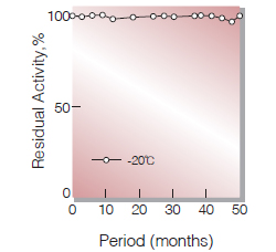
Fig.1. Stability (Powder form)
(kept under dry conditions)
-
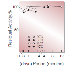
Fig.2. Stability (Powder form)
(kept under dry conditions)
-
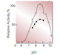
Fig.3. pH-Activity
The enzyme reaction was carried out with NADH(◯)or NADPH(●).buffer used: pH6.0-7.5, 10mM Veronal-acetate;pH7.5-9.0, 33mM Tris-HCI;pH9.2-10.2, 33mM NH4OH-NH4OH
-
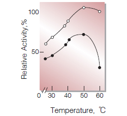
Fig.4. Temperature activity
The enzme reaction was carried out with NADH(◯)or NADPH(●).
20mM Tris-HCI buffer,pH8.5 -
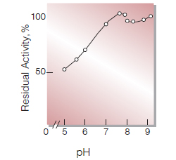
Fig.5. pH-Stability
30℃,3hr-treatment with the following buffer solution: pH6.0-7.5, 10mM Veronalacetate; pH7.5-9.0, 33mM Tris-HCI
-
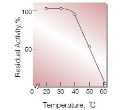
Fig.6. Thermal stability
30min-treatment with 0.2M Tris-HCI buffer,pH7.5 enzyme concentration:40U/ml
活性測定法(Japanese)
1. 原理
DCPIP(2,6-dichlorophenol-indophenol)の還元量を600nmの吸光度の変化で測定する。
2.定義
下記条件下で1分間に600nmの吸光度を1.0減少させる酵素量を1単位(U)とする。
3.試薬
- 0.2M Tris-HCl緩衝液, pH7.5
- 6.0mM NADH水溶液(用時調製,氷冷保存)
- 1.2mM DCPIP水溶液〔3.9mgのDCPIP・2H2O(MW=326.11)を10mlの蒸留水で溶解する〕(用時調製)
酵素溶液:酵素標品を予め氷冷した試薬Aに溶解し(約1.0%溶液),予め氷冷した0.1%牛血清アルブミン(BSA)を含む試薬Aで0.4〜0.8U/mlに希釈し,氷冷保存する。
4.手順
1.下記反応液をキュベット(d=1.0cm)に採り,25℃で約5分間予備加温する。
| 2.4ml | 蒸留水 | (A) |
| 0.3ml | Tris-HCl | (B) |
| 0.1ml | NADH水溶液 | (C) |
2.酵素溶液,DCPIP水溶液(C)各0.1mlをこの順序で添加し,ゆるやかにかつ速やかに混和後,水を対照に25℃に制御された分光光度計で600nmの吸光度変化を2〜3分間記録し,その初期直線部分より1分間当りの吸光度変化を求める(ΔODtest)。
3.盲検は反応混液1に酵素希釈液(0.1%BSAを含む試薬A),DCPIP水溶液(C)各0.1mlを加え,上記同様に操作を行って1分間当りの吸光度変化を求める(ΔODblank)。
5.計算式
U/ml =
-
ΔOD/min (ΔOD test−ΔOD blank)×希釈倍率
1.0×0.1(ml)
| = ΔOD/min×10×希釈倍率 | |
| U/mg | =U/ml×1/C |
| 1.0 | : 活性定義に基いて定められた600nmにおける単位吸光度 |
| C | : 溶解時の酵素濃度(c mg/ml) |
CONTACT
お問い合わせ-
各種製品に関するご質問・ご相談はこちらよりお問い合わせください。