-
HXK-311
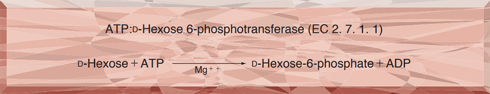
HEXOKINASE from Microorganism
PREPARATION and SPECIFICATION
| Appearance | White amorphous powder, lyophilized | |
|---|---|---|
| Activity | GradeⅢ 150U/mg-solid or more | |
| Contaminants | Phosphoglucose isomerase | ≤1.0×10⁻¹% |
| 6-Phosphogluconate dehydrogenase | ≤1.0×10⁻²% | |
| Glucose-6-phosphate dehydrogenase | ≤1.0×10⁻²% | |
| Myokinase | ≤1.0×10⁻²% | |
| Glutathione reductase | ≤5.0×10⁻¹% | |
PROPERTIES
| Stability | Stable at −20℃ for at least one year(Fig.1) |
|---|---|
| Molecular weight | approx. 82,000 (by gel filtration) |
| Isoelectric point | 4.1±0.1 |
| Michaelis constants | 2.3×10⁻⁴M (D-Glucose), 7.7×10⁻⁵M (ATP) |
| Inhibitors | Metal ions, p-chloromercuribenzoate, iodoacetamide, SDS, etc |
| Optimum pH | 8.0−9.0(Fig.2) |
| Optimum temperature | 50℃(Fig.3) |
| pH Stability | pH 4.0−9.0 (25℃, 20hr)(Fig.4) |
| Thermal stability | below 45℃ (pH 7.0, 30min)(Fig.5) |
| Substrate specificity | (Table 1) |
| Effect of various chemicals | (Table 2) |
APPLICATIONS
The enzyme is useful for enzymatic determination of glucose, adenosine-5'-triphosphate (ATP) and creatine phosphokinase when coupled with glucose-6-phosphate dehydrogenase (=G-6-PDH, G6D311, G6D-321).
ASSAY
Principle


The appearance of NADH is measured at 340nm by spectrophotometry.
Unit definition
One unit causes the formation of one micromole of NADH per minute under the conditions described below.
Method
Reagents
| A. Tris-HCl buffer, pH 8.0 | 50mM, containing 13.3mM MgCl2 | |
|---|---|---|
| B. Glucose solution | 0.67M in Tris-HCI buffer solution (A) (The solution Should be keep at roomtemperature at least for 1 hour before use) | |
| C. ATP solution | 16.5mM in Tris-HCl buffer solution (A) (Should be prepared fresh) | |
| D. NAD+ solution | 6.8mM in Tris-HCl buffer solution (A) (Should be prepared fresh) | |
| E. G-6-PDH solution | 300U/ml (Dilute with Tris-HCl buffer solution (A) and store on ice) | |
| F. Enzyme diluent | Tris-HCl buffer solution (A) contg. 0.1% of bovine serum albumin | |
Procedure
1. Prepare the following reactin mixture in a cuvette (d=1.0cm) and equilibrate at 30℃ for about 5 minutes.
| 2.30ml | Tris-HCl buffer solution | (A) |
| 0.50ml | Glucose solution | (B) |
| 0.10ml | ATP solution | (C) |
| 0.10ml | NAD+ solution | (D) |
| 0.01ml | G-6-PDH solution | (E) |
| Concentration in assay mixture | |
|---|---|
| Tris-HCl buffer | 50 mM |
| Glucose | 0.11 M |
| ATP | 0.53 mM |
| NAD+ | 0.22 mM |
| MgCl2 | 13 mM |
| BSA | 3.2μg/ml |
| G-6-PDH | ca.1.0 U/ml |
2. Add 0.1ml of the enzyme solution* and mix by gentle inversion.
3. Record the increase of optical density at 340nm against water for 4 to 5 minutes in a spectrophotometer thermostated at 30℃ and calculate theΔOD per minute from the initial portion of the curve (ΔOD test).
At the same time, measure the blank rate (ΔOD blank) by the same method as the test except the enzyme diluent (F) is added instead of the enzyme solution.
*Dissolve the enzyme preparation on ice-cold enzyme diluent (F) and dilute to 0.1−0.3U/ml with the same buffer, immediately before assay.
Calculation
Activity can be calculated by using the following formula :
Volume activity (U/ml) =
-
ΔOD/min (OD test−OD blank)×Vt×df
6.22×1.0×Vs
= ΔOD/min×5.0×df
Weight activity (U/mg) = (U/ml)×1/C
| Vt | : Total volume (3.11ml) |
| Vs | : Sample volume (0.1ml) |
| 1.0 | : Light path length (cm) |
| 6.22 | : Millimolar extinction coefficient of NADH (cm2/micromole) |
| df | : Dilution factor |
| C | : Enzyme concentration in dissolution (c mg/ml) |
Table 1. Substrate specificity of Hexokinase
[Pyruvate kinase-Lactate dehydrogenase system with 0.1M Tris-HCl buffer, pH 7.5]
-
Substrate(100mM) Relative activity(%) D-Glucose 100 D-Fructose 140 D-Mannose 52 2-Deoxy-D-glucose 91 -
Substrate(100mM) Relative activity(%) D-Galactose 0 D-Xylose 2 D-Glucosamine 58
Table 2. Effect of Various Chemicals on Hexokinase
[The enzyme dissolved in 50mM K-phosphate buffer, pH 6.5 (5U/ml) contg. 0.1% bovine serum albumin was incubated with each chemical at 30℃ for 1hr.]
-
Chemical Concn.(mM) Residual
activity(%)None - 100 Metal salt AgNO3 2.0 0 BaCl2 2.0 99 CaCl2 2.0 98 CdCl2 2.0 85 CoCl2 2.0 85 CuSO4 2.0 25 FeCl3 2.0 28 FeSO4 2.0 80 HgCl2 2.0 0 MgCl2 2.0 98 MnCl2 2.0 100 NiCl2 2.0 100 Pb(OAc)2 2.0 98 Zn(OAc)2 2.0 98 ZnSO4 2.0 99 NaF 20.0 101 NaN3 20.0 102 -
Chemical Concn.(mM) Residual
activity(%)PCMB 2.0 0 MIA 2.0 80 IAA 2.0 7 EDTA 5.0 103 (NH4)2SO4 20.0 104 Borate 20.0 102 o-Phenanthroline 2.0 101 α,α′-Dipyridyl 2.0 102 Urea 2.0 104 Guanidine 2.0 103 Hydroxylamine 2.0 104 Na-cholate 1.0% 102 Triton X-100 1.0% 105 Brij 35 1.0% 0 SDS 0.1% 25 Tween 20 0.1% 101 Span 20 0.1% 106 DAC 0.1% 101
Ac, CH3CO; NEM, N-Ethylmaleimide; PCMB, p-Chloromercuribenzoate; MIA, Monoiodoacetate; EDTA, Ethylenediaminetetraacetate; IAA, Iodoacetamide; SDS, Sodium dodecyl sulfate; DAC, Dimethylbenzylalkylammonium chloride.
-
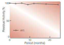
Fig.1. Stability (Powder form)
(kept under dry conditions)
-
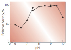
Fig.2. pH-Activity
30℃ in the 50mM buffer solution: pH6.2-7.5, PIPES-NaOH: pH7.5-9.0, Tris-HCI: pH9.0-10.0, Glycine-NaOH
-
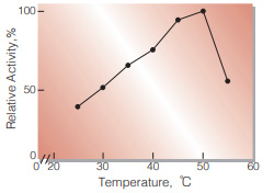
Fig.3. Temperature activity
(in 50mM Tris-HCI buffer,pH8.0)
-
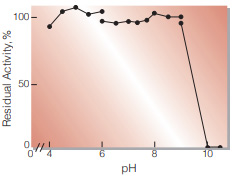
Fig.4. pH-Stability
25℃, 20hr-treatment in the 0.1M buffer solution: pH4.0-8.0,Acetate-NaOH;pH6.0-8.0, K-phosphate;pH7.5-9.0,Tris-HCl;pH9.0-10.5, Glycine-NaOH enzyme concn.: ca.10U/ml
-
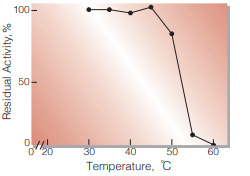
Fig.5. Thermal stability
30min-treatment with 50mM K-phosphate buffer, pH7.0, containing 0.1% bovine serum albumin enzyme concn.: ca.5U/ml
活性測定法(Japanese)
1. 原理
NADHの生成量を340nmの吸光度の変化で測定する。
2.定義
下記条件下で1分間に1マイクロモルのNADHを生成する酵素量を1単位(U)とする。
3.試薬
- 50mM Tris-HCl緩衝液,pH8.0 (13.3mM MgCl2を含む)
- 0.67Mグルコース緩衝液(試薬Aで溶解)(溶解後,室温に少くとも1時間保管したものを使用する)
- 16.5mM ATP溶液(試薬Aで溶解)(用時調製)
- 6.8mM NAD+溶液(試薬Aで溶解)(用時調製)
- G-6-PDH溶液(300U/mgに試薬Aで希釈し氷冷保存する)
酵素溶液:酵素標品を予め氷冷した0.1%牛血清アルブミン(BSA)を含む試薬Aの緩衝液で溶解し,同溶液で0.1〜0.3U/mgに希釈して氷冷保存する。
4.手順
1.下記反応混液をキュベット(d=1.0cm)に調製し,30℃で約5分間予備加温する。
| 2.30ml | Tris-HCl緩衝液 | (A) |
| 0.50ml | グルコース溶液 | (B) |
| 0.10ml | ATP溶液 | (C) |
| 0.10ml | NAD+水溶液 | (D) |
| 0.01ml | G-6-PDH溶液 | (E) |
2.酵素溶液を0.1mgを添加し,ゆるやかに混和後,水を対照に30℃に制御された分光光度計で,340nmの吸光度変化を4〜5分間記録し,その初期直線部分から1分間当りの吸光度変化を求める(ΔOD test)。
3.盲検は反応混液①に酵素溶液の代りに酵素希釈液(0.1%BSAを含む試薬A)を0.1mg加え,上記同様に操作を行って1分間当りの吸光度変化を求める。(ΔODblank)。
5.計算式
U/ml =
-
ΔOD/min (OD test−OD blank)×3.11(mg)×希釈倍率
6.22×1.0×0.1(mg)
| = ΔOD/min×5.0×希釈倍率 | |
| U/mg | = U/ml×1/C |
| 6.22 | : NADHのミリモル分子吸光係数(cm2/micromole) |
| 1.0 | : 光路長(cm) |
| C | : 溶解時の酵素濃度(c mg/ml) |
CONTACT
お問い合わせ-
各種製品に関するご質問・ご相談はこちらよりお問い合わせください。