LED-201
LEUCINE DEHYDROGENASE from Bacillus sp.1)
PREPARATION and SPECIFICATION
| Appearance | White amorphous powder, lyophilized | |
|---|---|---|
| Activity | GradeⅡ 20U/mg-solid or more (containing approx. 70% of stabilizers) |
|
| Contaminants | Leucylpeptide decomposing enzymes | |
| (Leu-Val) | ≤1.0×10⁻²% | |
| (Leu-Gly-Gly) | ≤1.0×10⁻²% | |
| NADH oxidase | ≤1.0×10⁻²% | |
| Stabilizers | 2-Mercaptoethanol, L-cysteine, dithiothreitol, ethylenediaminetetraacetate | |
PROPERTIES
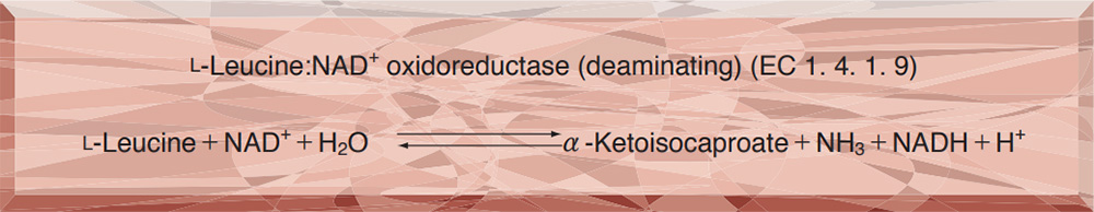
| Stability | Stable at −20℃ for at least one year(Fig.1) |
|---|---|
| Molecular weight2) | 245,000 |
| Michaelis constants2) | 1.0×10⁻³M (L-Leucine), 3.9×10⁻⁴M (NAD+), 3.5×10⁻⁵M (NADH), 3.1×10⁻⁴M [α-Ketoisocaproate (α-KⅠC)], 2.0×10⁻¹M (NH3) |
| Structure 2) | 6 subunits per enzyme molecule |
| Inhibitors 2) | Na2S, Hg++, Cu++, Co++, Mg++, p-chloromercuribenzoate |
| Optimum pH | 10.5−10.8 (L-Leu→α-KⅠC), 9.4 (α-KⅠC→L-Leu)(Fig.3) |
| Optimum temperature | above 70℃(Fig.4) |
| pH Stability | pH 5.5−10.5 (25℃, 20hr)(Fig.5) |
| Thermal stability | below 60℃ (pH 6.9, 10min)(Fig.6) |
| Substrate specificity | (Table 1) |
APPLICATIONS
This enzyme is useful for enzyme determination of L-leucine and the activity of leucine amino-peptidase.
ASSAY
Principle

The appearance of NADH is measured at 340nm by spectrophotometry.
Unit definition
One unit causes the formation of one micromole of NADH per minute under the conditions described below.
Method
Reagents
| A. L-Leucine solution | 0mM L-leucine in 0.2M glycine-KCl-KOH buffer, pH 10.5 (Prepare freshly) | |
|---|---|---|
| B. NAD+ solution | 12.5mM (Should be prepared fresh) | |
| C. Enzyme diluent | 25mM K-phosphate buffer, pH 7.2 | |
Procedure
1. Prepare the following reaction mixture in a cuvette (d=1.0cm) and equilibrate at 37℃ for about 5 minutes.
| 3.0ml | Substrate solution | (A) |
| 0.3ml | NAD+ solution | (B) |
| Concentration in assay mixture | |
|---|---|
| Glycine buffer | 0.18 M |
| L-Leucine | 18 mM |
| NAD+ | 1.1mM |
2. Add 0.05ml of the enzyme solution* and mix by gentle inversion.
3. Record the increase in optical density at 340nm against water for 2 to 3 minutes in a spectrophotometer thermostated at 37℃, and calculate the ΔOD per minute from the initial linear portion of the curve (ΔOD test).
At the same time, measure the blank rate (ΔOD blank) by using the same method as the test except that the enzyme diluent (C) is added instead of the enzyme solution.
*Dissolve the enzyme preparation in ice-cold enzyme diluent (C) (ca. 5mg/ml) and dilute to 0.25−0.33U/ml with the same buffer, immediately before assay.
Calculation
Activity can be calculated by using the following formula :
Volume activity (U/ml) =
-
ΔOD/min (ΔOD test−ΔOD blank)×Vt×df
6.22×1.0×Vs
= ΔOD/min×10.77×df
Weight activity (U/mg) = (U/ml)×1/C
| Vt | : Total volume (3.35ml) |
| Vs | : Sample volume (0.05ml) |
| 6.22 | : Millimolar extinction coefficient of NADH (㎠/micromole) |
| 1.0 | : Light path length (cm) |
| df | : Dilution factor |
| C | : Enzyme concentration in dissolution (c mg/ml) |
REFERENCES
1) K.Soda et al.; Biochem.Biophys.Res.Commun., 44, 931 (1971).
2) T.Ohshima et al.; J.Biol.Chem., 253, 5719 (1978).
Table 1. Substrate Specificity of Leucine dehydrogenase2)
-
Substrate(10mM) Relative activity(%) L-Leucine 100 L-Valine 74 L-Isoleucine 58 L-Norvaline 41 L-Norleucine 10 L-Methionine 0.6 L-Cysteine 0.3 Inert:L-Ala, L-Glu, L-Thr, L-Ser
Gly, L-Phe, L-Lys,
L-Arg, D-Leu, D-Val, D-Ile -
Substrate(10mM) Relative activity(%) α-Ketoisocaproate 100 α-Ketoisovalerate 126 α-Ketovalerate 76 α-Ketobutyrate 57 α-Ketocaproate 46 Inert:Pyruvate,α-Ketoglutarate,
Phenylpyruvate, Oxaloacetate,
Glyoxylate
-
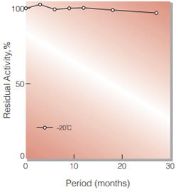
Fig.1. Stability (Powder form)
(kept under dry conditions)
-
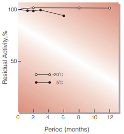
Fig.2. Stability (Powder form)
(kept under dry conditions)
-
Fig.3. pH-Activity
○̶̶○ :α-KIC→Leu in 1M ammonium buffer ●̶̶● :Leu→α-KIC in 0.2M glycineKOH buffer -
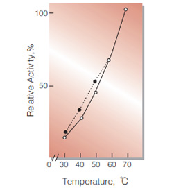
Fig.4. Temperature activity
○̶̶○ :α-KIC→Leu in 1.0M ammonium buffer pH9.5 ●̶̶● :Leu→α-KIC in 0.2M glycine-KOH bufferpH10.5 -
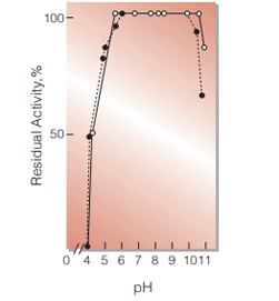
Fig.5. pH-Stability
25℃,20hr-treatment with 50mM buffer solution:pH4.0-6.0,acetate; pH6.0-8.5,phosphate;pH9.0-11.0, carbonate:
○̶̶○ :with 0.01% mercaptoethanol ●̶̶● :without mercaptoethanol -
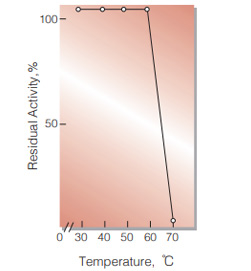
Fig.6. Thermal stability
10min-treatment with 50mM phosphate buffer,pH6.9
活性測定法(Japanese)
1. 原理
NADHの生成量を340nmの吸光度の変化で測定する。
2.定義
下記条件下で1分間に1マイクロモルのNADHを生成する酵素量を1単位(U)とする。
3.試薬
- 20mM L-ロイシン溶液〔L-ロイシンを20mMになるように0.2Mグリシン-KCl-KOH緩衝液(pH10.5)に溶解する〕(用時調製)
- 12.5mM NAD+水溶液(用時調製)
酵素溶液:酵素標品を予め氷冷した25mM K-リン酸緩衝液,pH7.2で溶解(約5mg/ml)し,分析直前に同緩衝液で0.25〜0.33U/mlに希釈する。
4.手順
1.下記反応混液をキュベット(d=1.0cm)に調製し,37℃で約5分間予備加温する。
| 3.0ml | 基質溶液 | (A) |
| 0.3ml | NAD+水溶液 | (B) |
2.酵素溶液0.05mlを添加し,ゆるやかに混和後,水を対照に37℃に制御された分光光度計で340nmの吸光度変化を2〜3分間記録し,その初期直線部分から1分間当りの吸光度変化を求める(ΔOD test)。
4.盲検は反応混液①に酵素溶液の代りに酵素希釈液(25mM K-リン酸緩衝液,pH7.2)を0.05mlを加え,上記同様に操作を行って,1分間当りの吸光度変化を求める(ΔODblank)。
5.計算式
U/ml =
-
ΔOD/min (ΔOD test−ΔOD blank)×3.35(ml)×希釈倍率
6.22×1.0×0.05(ml)
| = ΔOD/min×10.77×希釈倍率 | |
| U/mg | = U/ml×1/C |
| 6.22 | : NADHのミリモル分子吸光係数(cm2/micromole) |
| 1.0 | : 光路長(cm) |
| C | : 溶解時の酵素濃度(c mg/ml) |
CONTACT
お問い合わせ-
各種製品に関するご質問・ご相談はこちらよりお問い合わせください。