PCO-302
PROTOCATECHUATE 3,4-DIOXYGENASE from Pseudomonas sp.
PREPARATION and SPECIFICATION
| Appearance | Light brown amorphous powder, lyophilized | |
|---|---|---|
| Activity | GradeⅢ 3.0 U/mg-solid or more | |
| Contaminants | NADPH oxidase | ≤1.0×10⁻¹% |
| Stabilizers | Sugars | |
PROPERTIES
| Stability | Stable at −20℃ |
|---|---|
| Molecular weight | approx. 600,000 (by gel filtration) |
| Michaelis constant | 1.85×10⁻⁵M ((Protocatechuate) |
| Structure | Protein with nonheme ion |
| Inhibitors | Ag+, Hg++ |
| Optimum pH | 9.0 (Fig.2) |
| Optimum temperature | 60−65℃ (Fig.3) |
| pH Stability | pH 6.0−9.5 (25℃, 72hr) (Fig.4) |
| Thermal stability | below 55℃ (pH 7.5, 1hr) (Fig.5) |
| Effect of various chemicals | (Table 1) |
APPLICATIONS
This enzyme is useful for enzymatic determination of choline esterase when coupled with p-hydroxybenzoate hydroxylase (HBH-311).
ASSAY
Principle

The disappearance of protocatechuate is measured at 290nm by spectrophotometry.
Unit definition
One unit causes the oxidation of one micromole of protocatechuate per minute under the conditions described below.
Method
Reagents
| A. Tris-acetate buffer, pH 7.5 | 50mM[Dissolve 6.1g of Tris (MW=121.14) in ca.800ml of H2O and, after adjusting pH to 7.5 at 25℃ with 0.2M acetic acid, fill up to 1,000ml with H2O.] |
|---|---|
| B. Protocatechuate acid solution | 0.4mM[Dissolve 6.16mg of protocatechuate in ca.80ml of buffer (A) and, after adjusting pH to 7.5 at 25℃ with 1.0N KOH, fill up to 100ml with buffer (A).](Should be prepared fresh) |
Procedure
1.Pipette 3.0ml of protocatechuate solution (B) into a cuvette (d=1.0cm) and equilibrate at 37℃ for about 5 minutes.
| Concentration in assay mixture | |
|---|---|
| Tris-acetate buffer | 50 mM |
| Protocatechuate | 0.39mM |
2.Add 0.05ml of the enzyme solution* and mix by gentle inversion.
3.Record the decrease in optical density at 290nm against water for 3 to 4 minutes in a spectrophotometer thermostated at 37℃, and calculate the ΔOD per minute from the initial linear portion of the curve (ΔOD test).
At the same time, measure the blank rate (ΔOD blank) by using the same method as the test except that the enzyme diluent (A) is added instead of the enzyme solution.
*Dissolve the enzyme preparation in ice-cold diluent (A) (1.0mg/ml or more) and dilute to 0.2−0.8U/ml with the same buffer, immediately before assay.
Calculation
Activity can be calculated by using the following formula :
Volume activity (U/ml) =
-
ΔOD/min (ΔOD test−ΔOD blank)×Vt×df
3.8×1.0×Vs
= ΔOD/min×16.1×df
Weight activity (U/mg) = (U/ml)×1/C
| Vt | : Total volume (3.05ml) |
| Vs | : Sample volume (0.05ml) |
| 3.8 | : Millimolar extinction coefficient of protocatechuate (cm2/micromole) |
| 1.0 | : Light path length (cm) |
| df | : Dilution factor |
| C | : Enzyme concentration in dissolution (c mg/ml) |
REFERENCES
1) H.Fujisawa and O.Hayashi; J.Biol.Chem., 243, 2673 (1968)
Table 1. Effect of Various Chemicals on Protocatechuate 3,4-dioxygenase
[The enzyme dissolved in 50mM Tris-Acetate buffer (5U/ml) was incubated with each chemical at 30℃ for 1hr.]
-
Chemical Concn.(mM) Residual
activity(%)None - 100 Metal salt 1.0 AgNO3 26 BaCl2 97 CaCl2 97 CoCl2 97 CuSO4 95 FeSO4 79 MgSO4 100 MnCl2 100 NiCl2 100 ZnCl2 95 -
Chemical Concn.(mM) Residual
activity(%)NaF 1.0 100 NaN3 1.0 98 EDTA 5.0 98 Borate 50 97 SDS 0.05% 101 Brij 35 0.10% 103 Tween 20 0.10% 100 Na-cholate 0.10% 101
EDTA, ethylenediaminetetraacetate; SDS, sodium dodecyl sulfate.
-
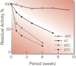
Fig.1. Stability (Powder form)
(kept under dry conditions)
-
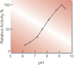
Fig.2. pH-Activity
(37℃ in 50mM Tris-Acetate buffer)
-
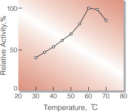
Fig.3. Temperature activity
(in 50mM K-phosphate buffer, pH 7.5)
-
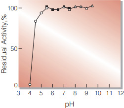
Fig.4. pH-Stability
25℃, 72hr-treatment with 50mM buffer solution: pH 4-5.5, Acetate; pH5.5-7.5 K-phosphate, pH7.0-9.5 Tris-acetate; Enzyme concentration: 5U/ml
-
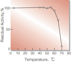
Fig.5. Thermal stability
1hr-treatment with 50mM Tris-acetate buffer, pH 7.5. Enzyme concentration: 5U/ml
活性測定法(Japanese)
1. 原理
プロトカテキュ酸の消失量を290nmの吸光度の変化で測定する。
2.定義
下記条件で1分間に1マイクロモルのプロトカテキュ酸が酸化される酵素量を1単位(U)とする。
3.試薬
- 50mM Tris-酢酸緩衝液, pH7.5〔6.1gのトリス (MW=121.14)を約800mlの蒸留水で溶解し, 0.2M酢酸でpH7.5(25℃)に調整後,蒸留水で 1,000mlとする〕
- 0.4mMプロトカテキュ酸溶液〔6.16mgのプロトカテ キュ酸を緩衝液(A)で溶解し,1N KOHでpH7.5 (25℃)に調整後,緩衝液(A)で100mlとする〕(用時調製)
酵素溶液:酵素標品を予め氷冷した緩衝液Aで溶解 (1.0mg/ml以上)し,分析直前に同緩衝液 で0.2〜0.8U/mlに希釈する。
4.手順
1.基質溶液(B)3.0mlをキュベット(d=1.0cm)に採り,37℃で約5分予備加温する。
2.酵素溶液0.05mlを添加し,ゆるやかに混和後,水を対照に37℃に制御された分光光度計で290nmの吸光 度変化を3〜4分間記録し,その初期直線部分から1分間当りの吸光度変化を求める(ΔOD test)。
3.盲検は基質溶液(B)に,酵素溶液の代わりに酵素希釈液(A)を0.05ml加え,上記同様に操作を行って,1分間当たりの吸光度変化を求める(ΔOD blank)。
5.計算式
U/ml =
-
ΔOD/min (ΔOD test−ΔOD blank)×3.05(ml)×希釈倍率
3.8×1.0×0.05(ml)
| = ΔOD/min×16.1×希釈倍率 | |
| U/mg | = U/ml×1/C |
| 3.8 | : プロトカテキュ酸のミリモル分子吸光係数(cm2/micromole) |
| 1.0 | : 光路長(cm) |
| C | : 溶解時の酵素濃度(c mg/ml) |
CONTACT
お問い合わせ-
各種製品に関するご質問・ご相談はこちらよりお問い合わせください。